Imaging a beating heart
Echocardiography (heart ultrasound) is a tool we use regularly to see the heart structure and function in real time in our pet patients. But when questions are complex or when surgical or interventional planning is being considered, a CT scan (also known as a CAT scan or computed tomography) adds crisp, 3D anatomy of the chambers, large vessels, and the coronary vessels that can be reconstructed for surgical planning.
At Boundary Bay Veterinary Specialty Hospital, we’ve added cardiac CT scans as another tool in our advanced diagnostic imaging services for select cases where extra detail

makes a meaningful difference. We asked Dr. Jennifer Gamracy (Residency Trained in Cardiology), and Deb Iampen (Manager of Diagnostic Imaging Services), to help us understand this new tool, how it works, and why it is an important addition to our hospital.
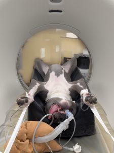
(Images for this post are of Dr. Gamracy’s dog, Odie, who helped us test the first scans of this new tool.)
What is the difference between cardiac CT and echocardiogram (echo)?
An echocardiogram (echo) is an ultrasound of the heart, which is routinely used to diagnose heart disease by identifying the size and function of the heart chambers, as well as the flow of blood through the heart. This can be done without anesthesia and often without sedation but relies on operator skill and a limited “window” of view of the heart through the lungs.
A CT scan is a form of non-invasive, advanced imaging that uses X-rays and a computer to create very thin “slices” (cross-sectional images) of the body that can be rebuilt into a 3D model and can be used for any part of a pet’s anatomy, including bones, lungs, chest, abdomen and nasal/sinus passages. A CT scan surpasses traditional X-rays by showing clearer, layered images without overlapping tissues.
A cardiac CT scan focuses on the heart and the major vessels around it—including the aorta, pulmonary artery, and coronary arteries (the small vessels that supply the heart muscle itself). The images are collected at specific time points in the heart’s beat, allowing still images of the beating heart. Because the patient must be very still throughout the scan, a cardiac CT scan requires general anesthesia for veterinary patients.
Learn more about our diagnostic imaging services, including ultrasound and MRI here.
How are cardiac CT images collected in pets?
Cardiac CT is different from other types of CT because of the use of electrocardiography (ECG) gating to create high-resolution, motion-free images. This is done using advanced computer systems that allow images to be collected at the moment in the pet’s heartbeat when there is little to no cardiac motion, and then the 3D images are reconstructed later from various directions and in multiple ways. When the heart is scanned throughout the entire ECG cycle, the image of the heart can also be reconstructed at any point in its beating cycle; systolic, diastolic, or anywhere in between. Functional data may also be obtained from the images.
To collect these images, the patient is deeply sedated (to prevent movement) and a breathing tube placed in their throat. The patient is placed on their back in a padded bed and connected to a special ECG. Medications are adjusted to keep the heart beating at a specific rate to get the clearest possible images. The padded bed is moved through the CT machine several times, as a special dye called “contrast” is injected into the patient’s vein to highlight the part of the heart we are most interested in.
After the images are collected, the patient wakes up and is given IV fluids to “flush” the contrast material from the bloodstream. In some cases, the patient may be moved directly into the surgery suite for surgical intervention based on the images collected.
What do cardiac CT images show?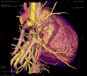
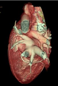
After the scan, we can generate volume-rendered images—true 3D views that can be rotated like a digital model. With these, we can:
- Identify the location of the coronary vessels (blood supply to the heart)
- Study the shapes of the heart chambers and how the great vessels (aorta, pulmonary artery) connect
- Explore abnormalities in the way the heart has formed (congenital heart diseases) and plan for surgical correction
Why is Cardiac CT an important new tool for evaluating the heart?
Although echocardiogram remains the best tool for day-to-day evaluation of the heart, our cardiology team can now use cardiac CT scans for cases where we need some extra information. It’s especially helpful when:
- Congenital abnormalities in the formation of the blood vessels away from the usual echocardiogram “window” is suspected
- Interventional or surgical decisions require detailed mapping of arteries and great vessels
- The precise location of a cardiac tumor needs to be identified for targeted radiation or surgical intervention
By pairing echocardiography’s live motion and flow information with cardiac CT’s high-definition 3D anatomy, we can give families clearer answers, plan procedures more definitively, and tailor treatment for pets who need that next level of detail.
How do I get a referral to Boundary Bay Veterinary Specialty Hospital’s Cardiology team?
There are two ways to get an appointment for specialty care at our hospital:
- From your primary veterinarian – A referral from your primary care vet ensures the proper transfer of medical information. It is beneficial to your pet and the specialty veterinarian to have all relevant medical information and will help your pet receive appropriate care. Once your veterinarian has sent a referral, we will contact you to set up an appointment. If you have not heard from us within 72 hours of your veterinarian sending a referral, please contact a client care representative at 604-514-8383.
- Contact us directly – You are welcome to contact us directly without a referral if you believe your pet would benefit from specialty or behaviour care. However, we believe strongly in working hand-in-hand with your family veterinarian to provide comprehensive care, and if it is possible to provide records or a referral from your family vet, it is helpful. Please contact us at 604-514-8383 or at info@bbvsh.com.
Frequently Asked Questions About Cardiac CT for Pets
What is cardiac CT for pets?
Cardiac CT is a specialized type of CT scan that focuses on the heart and major blood vessels, including the aorta, pulmonary artery, and coronary arteries. It creates high-resolution, 3D images of the heart that can be reconstructed at specific points in the heartbeat to show detailed anatomy.
How is cardiac CT different from an echocardiogram?
An echocardiogram (echo) is an ultrasound that evaluates heart size, function, and blood flow in real time and often does not require sedation or anesthesia. Cardiac CT, on the other hand, uses X-rays and advanced computer systems to create crisp, layered, 3D images of the heart and surrounding vessels. Cardiac CT requires general anesthesia because the patient must remain completely still during the scan.
When is cardiac CT used instead of echocardiography?
Echocardiography remains the primary tool for routine heart evaluation. Cardiac CT is used when more detailed anatomical information is needed, particularly for complex cases, surgical or interventional planning, or when structures are outside the usual echocardiogram viewing window.
What parts of the heart can cardiac CT visualize?
Cardiac CT provides detailed images of the heart chambers, coronary vessels, aorta, pulmonary artery, and other major vessels. It allows clinicians to study how these structures connect and identify abnormalities in heart formation.
Why does cardiac CT require anesthesia for pets?
Cardiac CT requires the patient to remain completely still to obtain motion-free images of the beating heart. General anesthesia allows precise image collection at specific moments in the heartbeat using ECG gating.
How are cardiac CT images collected?
During a cardiac CT scan, the pet is deeply sedated, intubated, and placed on a padded bed connected to an ECG. Images are collected at moments when the heart has little to no movement. A contrast dye is injected through a vein to highlight specific heart structures while the CT machine scans the heart over several heartbeats.
What is ECG gating in cardiac CT?
ECG gating is a technique that synchronizes image collection with the pet’s heartbeat. It allows high-resolution images to be captured when the heart is momentarily still, resulting in clear, detailed images of the heart throughout its beating cycle.
What does contrast dye do during a cardiac CT scan?
The contrast dye highlights specific areas of the heart and blood vessels, allowing clinicians to better visualize structures such as coronary arteries and major vessels. After the scan, IV fluids are given to help flush the contrast from the bloodstream.
What information do cardiac CT images provide?
Cardiac CT images can be reconstructed into true 3D models that can be rotated and examined from all angles. These images help identify coronary vessel locations, assess heart chamber shape, study vessel connections, and evaluate congenital heart abnormalities.
How does cardiac CT help with surgical or interventional planning?
Cardiac CT provides detailed 3D anatomy that can be reconstructed for surgical planning. It helps clinicians map arteries and great vessels, identify the precise location of tumors, and plan targeted surgical or interventional procedures.
Can cardiac CT detect congenital heart disease in pets?
Yes. Cardiac CT allows detailed evaluation of abnormalities in how the heart and blood vessels formed. This information is especially useful for planning surgical correction of congenital heart diseases.
What happens after a cardiac CT scan?
After imaging is complete, the pet wakes from anesthesia and receives IV fluids to flush the contrast material. In some cases, patients may be transferred directly to surgery based on the CT findings.
Why is cardiac CT an important addition to cardiac care?
By combining echocardiography’s live motion and blood flow information with cardiac CT’s high-definition 3D anatomy, clinicians can provide clearer answers, make more definitive treatment plans, and tailor care for pets that need advanced diagnostic detail.
Where is cardiac CT available for pets?
Cardiac CT is available at Boundary Bay Veterinary Specialty Hospital as part of its advanced diagnostic imaging services for select cases where additional detail can make a meaningful difference.
How do I get a referral for cardiac CT or cardiology services?
Appointments can be made either through a referral from your primary veterinarian or by contacting Boundary Bay Veterinary Specialty Hospital directly. Referrals help ensure proper transfer of medical information and coordinated care.
Who should consider cardiac CT for their pet?
Pets with complex heart conditions, suspected congenital vessel abnormalities, cardiac tumors, or those needing detailed surgical or interventional planning may benefit from cardiac CT when recommended by a veterinary cardiology team.
